Finding a new or strange-looking spot on your skin can trigger immediate anxiety. You ask yourself: Is this mole normal, or could it be skin cancer? Most pigmented spots on the human body are completely benign melanocytic nevi - harmless collections of pigment-producing cells called melanocytes. Yet, because melanoma is the most aggressive form of cutaneous malignancy, recognizing subtle signs of abnormality at the earliest stage is one of the most vital health habits you can develop.
What Does a Normal Mole Actually Look Like?
A typical benign mole (also called a common nevus) generally exhibits predictable characteristics. In healthy adults, most common moles share distinct visual traits:
- Symmetry: If you divide the spot vertically or horizontally across its center, both halves are near mirror images.
- Uniform Color: Normal moles are typically homogeneous - a single shade of pink, tan, medium brown, or dark brown throughout.
- Defined Margins: The transition between the mole's border and surrounding normal skin is sharp, crisp, and clearly demarcated.
- Modest Size: Most benign moles measure between 1 mm and 5 mm across (smaller than the diameter of a standard pencil eraser).
- Stability: Normal moles do not rapidly change their shape, outline, or elevation over weeks or months.
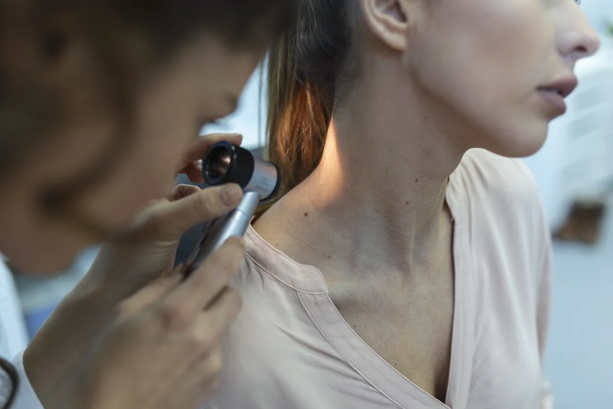
Benign vs. Malignant Comparison Chart
To help you evaluate your spots, dermatologists rely on comparative criteria. Here is how normal nevi differ from early malignant melanoma:
| Characteristic | Normal Benign Mole | Suspicious / Malignant Lesion |
|---|---|---|
| Symmetry | Round or oval, symmetrical halves | Asymmetrical, irregular contours |
| Border | Smooth, distinct, sharp perimeter | Scalloped, notched, frayed, or blurred |
| Color Distribution | Single uniform tan or brown tone | Mottled: black, brown, red, white, or slate blue |
| Diameter | Usually smaller than 6 mm | Frequently greater than 6 mm (can be smaller) |
| Behavior Over Time | Stable and static for years | Evolving: growing, changing color, or bleeding |
Atypical Moles (Dysplastic Nevi): The In-Between Category
Not every unusual mole is cancerous. Approximately 10% of fair-skinned individuals have dysplastic nevi - clinically atypical moles that share some visual features with melanoma (such as slightly irregular borders or mild color variation) but remain histologically non-malignant under biopsy. However, individuals with numerous atypical moles carry a statistically higher lifetime risk of developing melanoma elsewhere on their skin.
When Should You See a Board-Certified Dermatologist?
You should seek prompt medical evaluation if you notice any spot demonstrating the "Three S Rules":
- Spontaneous Bleeding or Oozing: A mole that bleeds without physical trauma or scratching.
- Sensory Symptoms: Persistent localized itching, tenderness, stinging, or pain.
- Sudden Evolution: Rapid enlargement, darkening, or surface changes over 4 to 8 weeks.
Remember: skin cancer is highly curable when detected in its earliest intraepidermal stages (Stage 0 Melanoma in situ). Developing a consistent habit of monthly photographic self-examinations provides the objective record your doctor needs to make accurate clinical decisions.
Monitor Your Skin & Moles Confidently
Spot changes before they escalate. Capture clinical-grade photo timelines, map spots on an interactive 3D body map, and share structured reports with your dermatologist.
Télécharger Gratuitement sur iOSFoire Aux Questions (FAQ)

Anna Brown, MSc
Lead Clinical WriterAnna is a medical health writer specializing in cutaneous oncology, dermoscopy, and AI dermatology. Her guides are verified against WHO, AAD, and peer-reviewed clinical literature.
View All 21 Articles by Anna Brown →Scientific References & Clinical Studies
- Skin Cancer Foundation: Melanoma Warning Signs — Skin Cancer Foundation Clinical Guidelines
- WHO Fact Sheets on Skin Cancer & Ultraviolet Radiation — World Health Organization (WHO)
- Atypical Dysplastic Nevi Clinical Overview — PubMed / National Institutes of Health (NIH)
